How is epilepsy diagnosed?
As we discussed earlier, epilepsy may be diagnosed after two unprovoked seizures occurring more than 24 hours apart, or after one unprovoked seizure if the risk of having another seizure is considered sufficiently high—at least 60% over the next 10 years. Certain epilepsy syndromes can also establish the diagnosis.
The first step, therefore, is to determine whether the event that occurred was actually an epileptic seizure. Many other conditions can sometimes resemble seizures, so understanding exactly what happened before, during and after the event is extremely important.
For this reason, we usually take a detailed account from both the patient and someone who witnessed the event, whenever possible. A video recording of the event, particularly when obtained safely on a mobile phone, can also be extremely useful in helping your doctor understand what happened.
Your doctor will then consider the nature of the event along with other clinical information—such as your medical history, examination, previous illnesses or injuries, family history and other factors that may help determine the likelihood of epilepsy and the risk of further seizures.
Depending on the circumstances, investigations may then be advised. These commonly include an EEG (electroencephalogram), MRI or other imaging of the brain, and sometimes blood tests or other investigations.
Importantly, there is no single test that can diagnose or rule out epilepsy in every person. The diagnosis is made by putting together the description of the event, your clinical history and examination, and the results of appropriate investigations.
What is an EEG and why do I need one?
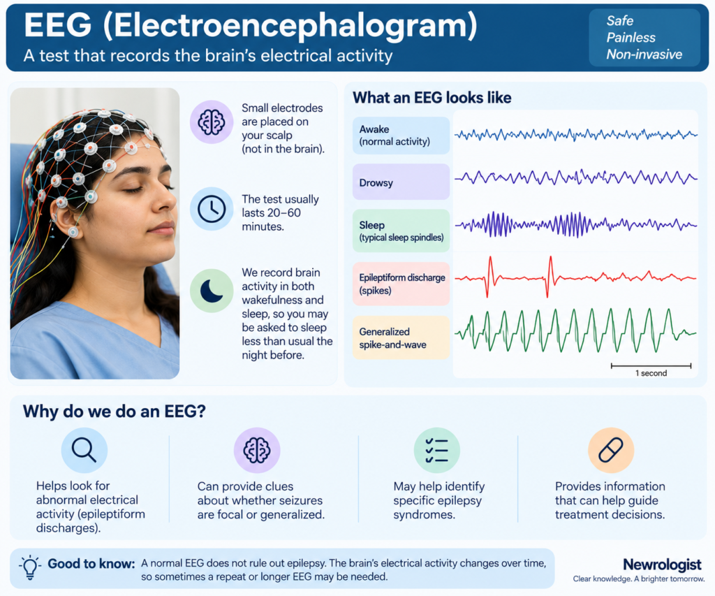
An electroencephalogram (EEG) is an investigation in which we record the electrical activity of your brain. A set of small electrodes is temporarily placed over your scalp to record this activity. A routine EEG commonly lasts around 20–60 minutes, although longer recordings may sometimes be required.
Ideally, we would like to record the brain during both wakefulness and sleep, because some abnormalities become easier to detect during drowsiness or sleep. For this reason, you may sometimes be asked to sleep less than usual the night before your EEG so that you are more likely to fall asleep during the recording.
Our brain normally produces characteristic patterns of electrical activity, which change depending on whether we are awake, drowsy or asleep. During an EEG, we examine these normal patterns as well as look for abnormalities.
Occasionally, a seizure may occur during the recording, allowing us to see the electrical changes associated with the seizure. This can be particularly informative when the EEG is recorded together with video.
More commonly, however, we look for abnormalities that can occur between seizures, known as epileptiform discharges. You may hear terms such as spikes, sharp waves or spike-and-wave discharges. Their location and pattern can provide important clues about whether seizures are likely to be focal or generalized, and in focal epilepsy may sometimes provide information about the brain region involved.
Certain characteristic EEG patterns can also help us identify particular epilepsy syndromes. All of this information—when interpreted together with your history and other investigations—can help your neurologist classify the epilepsy, establish the diagnosis in appropriate circumstances and choose the most suitable treatment.
My EEG was normal. Does that mean I don’t have epilepsy?
No. A normal EEG does not rule out epilepsy.
During a routine EEG, we record the brain’s electrical activity for only a relatively short period of time. A person with epilepsy does not continuously have abnormal electrical discharges. It is therefore entirely possible that no epileptiform abnormality occurs during the period in which the EEG is being recorded.
In fact, a routine EEG may detect epileptiform abnormalities in only around half of people with epilepsy. The chances of detecting these abnormalities can be increased in several ways.
Repeating the EEG can increase its yield, as an abnormality that was not seen during the first recording may appear during a subsequent one. Recording sleep as well as wakefulness is also useful because epileptiform abnormalities become more apparent during sleep in many forms of epilepsy. This is one reason why your doctor may request a sleep-deprived EEG.
In selected patients, we may perform prolonged video-EEG monitoring, where both the EEG and a synchronized video of the person are recorded continuously for a much longer period—from several hours to sometimes several days. This allows us to try to capture the person’s actual episodes and determine what happens in the brain at the same time. It is particularly useful when the diagnosis remains uncertain, when seizures continue despite treatment, or as part of the evaluation for epilepsy surgery.
So, a normal EEG does not mean that you do not have epilepsy. Epilepsy remains primarily a clinical diagnosis, with the EEG providing important supporting information.
On the other hand, characteristic epileptiform abnormalities on an EEG can strongly support a diagnosis of epilepsy, help determine the type of epilepsy, and provide information about the risk of further seizures.
Why do I need an MRI of the brain?
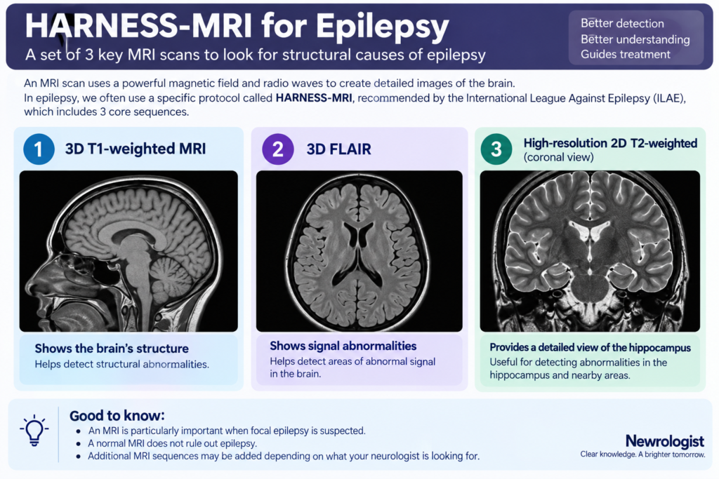
In most people in whom epilepsy is suspected or diagnosed, your doctor may advise an MRI scan of the brain. The purpose of the MRI is primarily to look for a structural abnormality in the brain that might be responsible for the seizures.
This is particularly important when we suspect focal epilepsy. As we discussed earlier, focal seizures begin within a particular network on one side of the brain. The MRI allows us to look carefully for structural abnormalities that could explain why seizures are arising from that region. These may include abnormalities that have been present since brain development, previous injuries or strokes, tumors, or other structural lesions.
An MRI can also be useful when it remains uncertain whether the epilepsy is focal or generalized. However, in some people with a very typical generalized epilepsy syndrome, where the clinical history and EEG findings are characteristic, your neurologist may decide that an MRI is not necessary.
Importantly, an epilepsy MRI is somewhat different from a routine MRI of the brain. The ILAE has recommended a standardized set of high-resolution MRI sequences called the HARNESS-MRI protocol, designed to improve the detection of structural abnormalities associated with epilepsy.
Your neurologist may therefore specifically request an epilepsy-protocol MRI, particularly when focal epilepsy is suspected or when a previous routine MRI has not adequately answered the clinical question.
And just as with EEG, a normal MRI does not rule out epilepsy. Many people with epilepsy have no structural abnormality visible on currently available MRI scans.
Leave a Reply